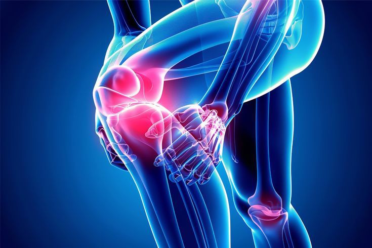
Medial Collateral Ligament
What Is an MCL Injury?
A Medial Collateral Ligament (MCL) injury is one of the most common ligament injuries affecting the knee. The MCL is a strong band of connective tissue located on the inner (medial) side of the knee. It stabilizes the knee by preventing excessive inward movement (valgus stress) and supports normal knee function during walking, running, and sports activities.
MCL injuries range from mild stretching of the ligament to complete tears. They are particularly common among athletes participating in contact sports but may also occur after falls, twisting injuries, or road traffic accidents.
Anatomy of the MCL
The MCL extends from the medial femoral epicondyle to the medial aspect of the tibia. It consists of:
- Superficial MCL
- Deep MCL
These structures work together to resist valgus stress and rotational forces while maintaining medial knee stability.
Etiology (Causes)
The most common mechanism of MCL injury is valgus stress, in which a force pushes the knee inward while the lower leg moves outward, overstretching or tearing the ligament.
Contact Injury (Most Common)
Contact injuries are more common than non contact injuries and usually occur due to:
- Direct blow to the lateral (outer) side of the knee
- Football injuries
- Rugby injuries
- Hockey injuries
- Motor vehicle accidents
- Falls
A direct blow to the outside of the knee produces a valgus force, causing injury to the MCL.
Non Contact Injury
Non contact injuries are less common and usually occur during:
- Sudden twisting
- Pivoting
- Rapid change in direction
- Landing awkwardly after a jump
These injuries often result in Grade I or Grade II (incomplete) ligament tears.
Risk Factors
People are at increased risk if they:
- Participate in contact sports
- Have weak quadriceps or hamstrings
- Have had previous knee injuries
- Perform inadequate warm-up before exercise
- Use poor sporting techniques
Associated Injuries and Conditions
Anterior Cruciate Ligament (ACL) Tear
MCL injuries are commonly associated with ACL tears.
Important facts:
- Combined ACL MCL injury is the most common multiple-ligament injury of the knee.
- Hemarthrosis (blood inside the knee joint) strongly suggests an associated ACL injury.
- The risk of ACL injury increases with the severity of the MCL injury:
- Grade III > Grade II > Grade I
Meniscus Tear
The meniscus may also be injured.
- Medial meniscus tears are more common than lateral meniscus tears.
- Up to 5% of isolated MCL injuries are associated with meniscal tears.
- Patients may complain of locking, catching, or clicking sensations.
Pellegrini Stieda Syndrome
A chronic complication of MCL injury characterized by:
- Calcification at the femoral attachment of the MCL
- Chronic pain on the inner side of the knee
- Stiffness
- Reduced range of motion
This condition usually develops after chronic MCL injury or inadequate healing.
Symptoms
Patients commonly experience:
- Pain on the inner (medial) side of the knee
- Swelling
- Tenderness over the MCL
- Knee stiffness
- Bruising (ecchymosis)
- Difficulty walking
- Feeling that the knee “gives way”
- Reduced range of motion
- A “pop” at the time of injury
Classification
Grade I (Mild)
- Ligament stretched without tearing
- Mild pain
- Minimal swelling
- No instability
- Recovery: 1–3 weeks
Grade II (Moderate)
- Partial ligament tear
- Moderate pain and swelling
- Mild to moderate instability
- Difficulty walking
- Recovery: 3–6 weeks
Grade III (Severe)
- Complete ligament rupture
- Significant instability
- Severe pain and swelling
- Difficulty bearing weight
- Recovery: 6–12 weeks or longer
Clinical Examination
A complete examination includes:
Inspection
The healthcare provider looks for:
- Knee swelling (effusion)
- Bruising (ecchymosis)
- Deformity
Palpation
The examiner checks for:
- Tenderness along the medial aspect of the knee
- Pain over the MCL attachment
- Localized swelling
Range of Motion
Flexion and extension are assessed to identify movement limitations.
Valgus Stress Test
The valgus stress test is the most important clinical examination for diagnosing MCL injuries.
Technique
- Knee flexed to 30°
- Examiner stabilizes the thigh
- Valgus force applied to the lower leg
Interpretation
- Pain without laxity = Grade I
- Pain with mild laxity = Grade II
- Marked laxity without a firm endpoint = Grade III
Investigations
X ray
Performed to exclude fractures or bony avulsion injuries.
MRI
The gold standard for diagnosing MCL injuries. MRI identifies:
- Partial tears
- Complete tears
- ACL injuries
- Meniscal tears
- Cartilage injuries
Ultrasound
May be useful for evaluating superficial ligament injuries.
Treatment
Conservative Management
Most isolated MCL injuries heal without surgery.
Treatment includes:
- Rest
- Ice (15–20 minutes every 2–3 hours during the first 48 hours)
- Compression
- Elevation
- NSAIDs (when appropriate)
- Hinged knee brace
- Crutches if needed
- Early physiotherapy
Rehabilitation
Phase 1
- Reduce pain and swelling
- Gentle range of motion exercises
Phase 2
- Quadriceps strengthening
- Hamstring strengthening
- Hip strengthening
Phase 3
- Balance and proprioception exercises
- Functional training
Phase 4
- Running progression
- Agility drills
- Sport specific rehabilitation
- Return to play assessment
Surgical Management
Surgery may be required for:
- Complete Grade III tears with persistent instability
- Combined ACL MCL injuries
- Bony avulsion injuries
- Failure of conservative treatment
- Chronic medial knee instability
Procedures include:
- MCL repair
- MCL reconstruction using tendon grafts
Recovery Time
Grade Recovery Grade I 1–3 weeks Grade II 3–6 weeks Grade III 6–12 weeks or longer
Complications
Without appropriate treatment, complications may include:
- Chronic medial knee instability
- Persistent pain
- Knee stiffness
- Recurrent ligament injuries
- Early osteoarthritis
- Pellegrini Stieda syndrome
- Muscle weakness
Prevention
- Warm up before exercise.
- Strengthen the quadriceps, hamstrings, and hip muscles.
- Improve flexibility and balance.
- Wear appropriate footwear and protective sports equipment.
- Use proper sports techniques.
- Avoid sudden twisting movements when possible.
Frequently Asked Questions (FAQs)
Can an MCL tear heal without surgery?
Yes. Most isolated Grade I and Grade II tears, and many Grade III injuries, heal successfully with conservative treatment and rehabilitation.
What is the most common mechanism of MCL injury?
A valgus stress injury, usually caused by a direct blow to the outer side of the knee, is the most common mechanism.
Which injuries are commonly associated with MCL tears?
ACL tears and medial meniscus tears are the most common associated injuries.
What is the best imaging test for MCL injuries?
MRI is the preferred imaging modality because it accurately identifies the extent of ligament injury and associated knee damage.
Disclaimer: This article is intended for educational purposes only and should not replace professional medical advice, diagnosis, or treatment. Always consult a qualified healthcare provider for evaluation and management of knee injuries.
© aminarehman.com. All rights reserved.


Leave a Reply