Introduction
Coronary Artery Disease (CAD) is one of the leading causes of illness and death worldwide. It develops when the coronary arteries, which supply oxygen rich blood to the heart muscle, become narrowed or blocked by the gradual accumulation of fatty deposits known as atherosclerotic plaques. Reduced blood flow deprives the heart of oxygen, resulting in myocardial ischemia. If the blockage becomes complete, the affected heart muscle begins to die, causing a myocardial infarction (heart attack).
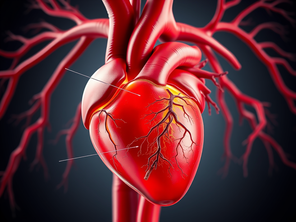
Coronary Artery Anatomy
The heart receives its blood supply through two main coronary arteries that originate from the ascending aorta.
Left Coronary Artery (LCA)
- Left Anterior Descending (LAD)
- Left Circumflex (LCX)
Right Coronary Artery (RCA)
Together, these vessels provide oxygen and nutrients to the myocardium, enabling the heart to pump efficiently.
Pathophysiology of Coronary Artery Disease
1. Endothelial Injury
The disease begins with damage to the endothelial lining of the coronary arteries. Common causes include hypertension, cigarette smoking, diabetes mellitus, elevated LDL cholesterol, obesity, chronic inflammation, and physical inactivity.
A damaged endothelium loses its protective function, allowing lipoproteins and inflammatory cells to penetrate the arterial wall.
2. Lipid Accumulation
Low density lipoprotein (LDL) cholesterol enters the arterial wall and undergoes oxidation. Oxidized LDL triggers an inflammatory response, attracting white blood cells and promoting further endothelial dysfunction.
3. Formation of Fatty Streaks
Monocytes migrate into the intima and differentiate into macrophages. These macrophages engulf oxidized LDL, becoming foam cells. Collections of foam cells form fatty streaks, representing the earliest visible stage of atherosclerosis.
4. Development of Atherosclerotic Plaques
As inflammation continues, smooth muscle cells migrate into the intima and produce collagen and extracellular matrix, forming a fibrous cap over a lipid rich core. Over time, calcium may also be deposited within the plaque, making the artery stiffer.
5. Progressive Narrowing of the Coronary Artery
As plaques enlarge, the arterial lumen gradually narrows, reducing coronary blood flow. During periods of increased oxygen demand, such as exercise or emotional stress, the narrowed artery cannot supply enough blood to the myocardium, resulting in stable angina.
6. Plaque Rupture and Thrombus Formation
Some plaques become unstable and rupture. The exposed plaque material activates platelets and the coagulation cascade, leading to thrombus formation. The blood clot may partially or completely obstruct the coronary artery.
7. Myocardial Infarction
Complete obstruction of a coronary artery interrupts blood flow to the myocardium. If ischemia persists for more than 20–30 minutes, irreversible myocardial cell death (necrosis) occurs, resulting in an acute myocardial infarction.
Risk Factors
Non Modifiable Factors
- Increasing age
- Male sex
- Postmenopausal status in women
- Family history of premature coronary artery disease
Modifiable Factors
- Smoking
- Hypertension
- Diabetes mellitus
- Dyslipidemia
- Obesity
- Sedentary lifestyle
- Unhealthy diet
- Chronic stress
Clinical Features
Patients may present with:
- Chest pain or pressure (angina)
- Shortness of breath
- Fatigue
- Pain radiating to the left arm, neck, jaw, shoulder, or back
- Nausea and vomiting
- Excessive sweating
- Dizziness or fainting
Symptoms vary according to the severity of coronary artery obstruction.
Diagnostic Evaluation
The diagnosis of CAD is established using a combination of clinical assessment and investigations.
Laboratory Tests
- Cardiac Troponin I or T
- CK MB
- Lipid Profile
- Blood Glucose
- HbA1c
Electrocardiography (ECG)
ECG helps detect myocardial ischemia or infarction through changes such as ST-segment depression, ST segment elevation, T-wave inversion, or pathological Q waves.
Imaging Studies
- Echocardiography
- Exercise Stress Test
- Coronary CT Angiography
- Coronary Angiography (Gold Standard)
Management
Lifestyle Modification
- Smoking cessation
- Healthy diet
- Regular physical exercise
- Weight reduction
- Blood pressure control
- Diabetes management
- Stress reduction
Pharmacological Treatment
- Antiplatelet agents
- Statins
- Nitrates
- Beta blockers
- ACE inhibitors or ARBs
- Calcium channel blockers (when indicated)
Revascularization
Patients with severe disease may require:
- Percutaneous Coronary Intervention (PCI) with balloon angioplasty and stent placement
- Coronary Artery Bypass Grafting (CABG)
Complications
- Myocardial infarction
- Heart failure
- Cardiac arrhythmias
- Cardiogenic shock
- Sudden cardiac death

Copyright © 2026 Amina Rehman. All Rights Reserved.

Leave a Reply