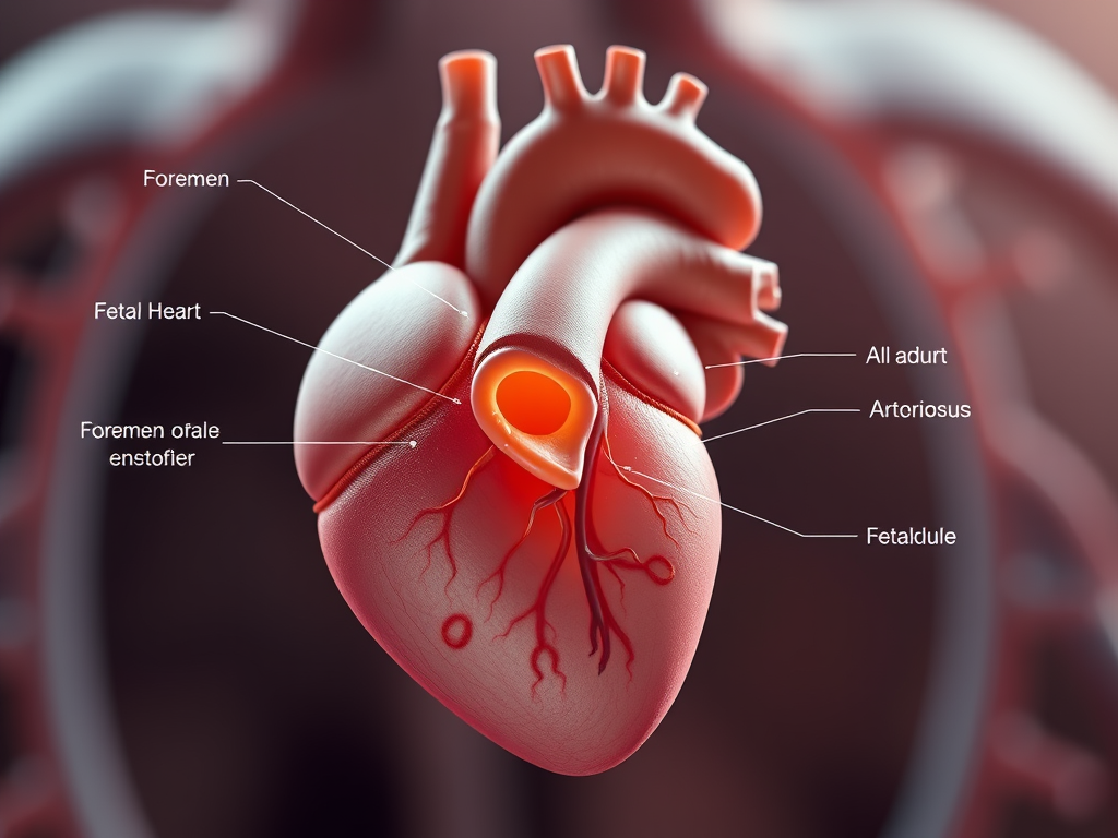
The fetal heart anatomy differs from an adult heart in several ways. Here are some key points:
- Foramen Ovale: In a fetus, there’s a small hole called the foramen ovale in the atrial septum (the wall between the two upper chambers of the heart). This hole allows blood to bypass the fetal lungs since oxygen is obtained from the mother’s blood via the placenta.
- Ductus Arteriosus: Another important structure is the ductus arteriosus, a blood vessel that connects the pulmonary artery and the aorta. It also helps divert blood away from the lungs and towards the rest of the body.
- High Pulmonary Pressure: Fetal blood pressure in the pulmonary artery is much higher than in the aorta because the lungs are not being used for oxygen exchange. This pressure difference keeps the foramen ovale and ductus arteriosus open.
- Small Size: The fetal heart is proportionally smaller compared to the body size and undergoes significant growth after birth.
- Specialized Shunts: These structures and circulation patterns gradually change after birth. This change occurs as the baby takes its first breaths. This action initiates the oxygenation of blood in the lungs. The foramen ovale and ductus arteriosus usually close within the first days to months of life, allowing the heart to function in the typical adult pattern.
The fetal heart undergoes significant developmental changes. These changes help it adapt to the unique environment in the womb. After birth, it transitions to its adult form and function.
CATEGORIES
fetus
Fetal heart
