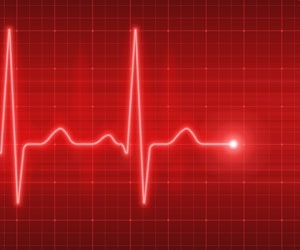
1. What is Bradycardia?
- Definition: Heart rate slower than normal.
- Number: Less than 60 beats per minute (bpm) in adults at rest.
- Important: Bradycardia can be normal (like in athletes during sleep) or abnormal (causing symptoms like dizziness).
Key point: A fit athlete at 45 bpm is fine. An elderly person at 50 bpm with fainting is not.
2. Why Does Heart Rate Slow Down?
The heart’s natural pacemaker is called the SA node. Bradycardia happens when:
- The SA node fires too slowly.
- The electrical signal gets blocked between the upper and lower chambers of the heart.
- The vagus nerve (which slows the heart) is overactive.
- Medicines or diseases suppress the heart rate.
3. Causes of Bradycardia
A. Normal (Physiological) Causes
- Athletes (athletic heart)
- Deep sleep
- Young healthy people
B. Medical (Pathological) Causes
- Sick sinus syndrome (SA node fails)
- Heart blocks (AV blocks)
- Heart attack (especially inferior wall MI)
- After heart surgery
Other causes:
- Medicines: Beta blockers, calcium channel blockers, digoxin, amiodarone
- Electrolyte problem: High potassium
- Underactive thyroid (hypothyroidism)
- Increased brain pressure (Cushing’s reflex)
- Low body temperature (hypothermia)
- Infections like Lyme disease
- Vagal maneuvers (coughing, bearing down to pass stool)
4. Symptoms (When Bradycardia Becomes a Problem)
If the heart beats too slowly to pump enough blood, these symptoms appear:
- Early stage: Tiredness, weakness, feeling lightheaded, almost fainting
- Moderate stage: Shortness of breath with activity, confusion
- Severe stage: Fainting, chest pain, low blood pressure, shock
Classic three symptoms: Fainting, fatigue, dizziness
No symptoms? Usually no treatment needed.
5. ECG Diagnosis of Bradycardia
ECG is the best test to diagnose bradycardia. It shows:
- The actual heart rate
- Where the rhythm is coming from
- Any block in electrical conduction
Step 1: Calculate Heart Rate on ECG
- If rhythm is regular: Divide 300 by the number of large squares between two R waves.
- Or count QRS complexes in 6 seconds and multiply by 10.
- Bradycardia = less than 60 bpm.
Step 2: Identify the Type of Bradycardia on ECG
A. Sinus Bradycardia
- ECG signs: Normal P wave before every QRS, normal PR interval, normal QRS shape, rate below 60.
- Causes: Athletes, vagal tone, medicines, low thyroid, inferior heart attack.
B. First Degree AV Block
- ECG signs: PR interval longer than 200 ms (one large square). Every P wave conducts to QRS.
- Outcome: Usually harmless.
C. Second Degree AV Block Type I (Mobitz I / Wenckebach)
- ECG signs: PR interval gets longer and longer until a QRS is dropped. Then the pattern repeats.
- Location: Usually in the AV node. Often benign.
D. Second Degree AV Block : Type II (Mobitz II)
- ECG signs: PR interval is constant, but suddenly a P wave is not followed by a QRS. No warning prolongation.
- Danger: This is more serious. Can suddenly progress to complete heart block.
E. Third Degree AV Block (Complete Heart Block)
- ECG signs: Atria and ventricles beat completely independently. P waves and QRS complexes have no relationship. Ventricular rate is very slow (30–50 bpm or even slower).
- QRS shape: Narrow (better) or wide (worse prognosis).
F. Junctional Bradycardia
- ECG signs: Rate below 60. No P wave, or an inverted P wave. Narrow QRS.
- Cause: SA node is suppressed (digoxin toxicity, inferior MI).
G. Escape Rhythms (Backup rhythms)
- Atrial escape: Rate 50–60, abnormal P wave.
- Junctional escape: Rate 40–60, no P wave.
- Ventricular escape: Rate 20–40, wide QRS this is the heart’s last resort.
6. Quick Reference Table for ECG Patterns
| Type | Rate | P Wave | PR Interval | QRS | Rhythm |
|---|---|---|---|---|---|
| Sinus brady | <60 | Normal, before each QRS | Normal | Narrow | Regular |
| 1° AV block | Any | Normal | >200 ms | Narrow | Regular |
| 2° Mobitz I | Usually <60 | Normal | Gets longer then drops | Narrow | Grouped beats |
| 2° Mobitz II | Often slow | Normal | Fixed, then drops | Narrow/wide | Irregular |
| 3° AV block | Atrial 60-100, Ventricular 30-50 | Normal | No relation | Narrow or wide | Regular but separate |
| Junctional brady | <60 | Absent or inverted | Short (<120) | Narrow | Regular |
7. Further Tests (After ECG)
- Holter monitor (24 hour recording) : for intermittent bradycardia.
- Echocardiogram : to check heart structure.
- Blood tests: Thyroid function, electrolytes, troponin (if heart attack suspected), digoxin level.
- Exercise stress test : if heart rate doesn’t increase properly with activity.
8. Treatment of Bradycardia
A. Asymptomatic (No symptoms)
- No treatment needed.
- Stop any offending medicine.
- Remove reversible causes.
B. Symptomatic Bradycardia (Guideline based treatment)
Immediate treatment (ABCs):
- Atropine 0.5 mg IV every 3-5 minutes (max 3 mg) : first drug of choice.
- Transcutaneous pacing (if atropine fails or patient is very unstable).
- Dopamine or epinephrine infusion (if pacing is not available).
Caution: Do not use atropine in complete heart block with wide QRS . it usually won’t work.
C. Long term Treatment
- Pacemaker implantation : definitive treatment for symptomatic sinus node dysfunction, Mobitz II, or complete heart block.
- Stop unnecessary heart slowing medicines.
9. Clinical Pearls (Important Tips)
- Athletes: Resting heart rate of 30–40 bpm and pauses of 2–3 seconds during sleep are normal.
- Inferior heart attack: Often causes first degree or Mobitz I block. Usually temporary and rarely needs a pacemaker.
- Sick sinus syndrome: ECG may show fast and slow rhythms alternating (tachy brady syndrome). Needs pacemaker plus medicines for fast rhythms.
- Medicine-induced: Always check for beta blockers, verapamil, diltiazem, digoxin, and amiodarone.
- Carotid sinus hypersensitivity: Massaging the neck can cause a pause of more than 3 seconds. Treat with a pacemaker.
10. Summary Table for Quick Review
| Parameter | Detail |
|---|---|
| Definition | Heart rate < 60 bpm in adults |
| Most common cause | Sinus bradycardia (physiologic or vagal) |
| Most dangerous ECG | 3° AV block with wide QRS escape |
| First ACLS drug | Atropine 0.5 mg IV |
| Definitive therapy | Pacemaker (if symptomatic and irreversible) |
| Always do first | ECG to identify rhythm and block type |
Copyright Notice: © 2025 amamedico. All rights reserved. Unauthorized reproduction is prohibited.

